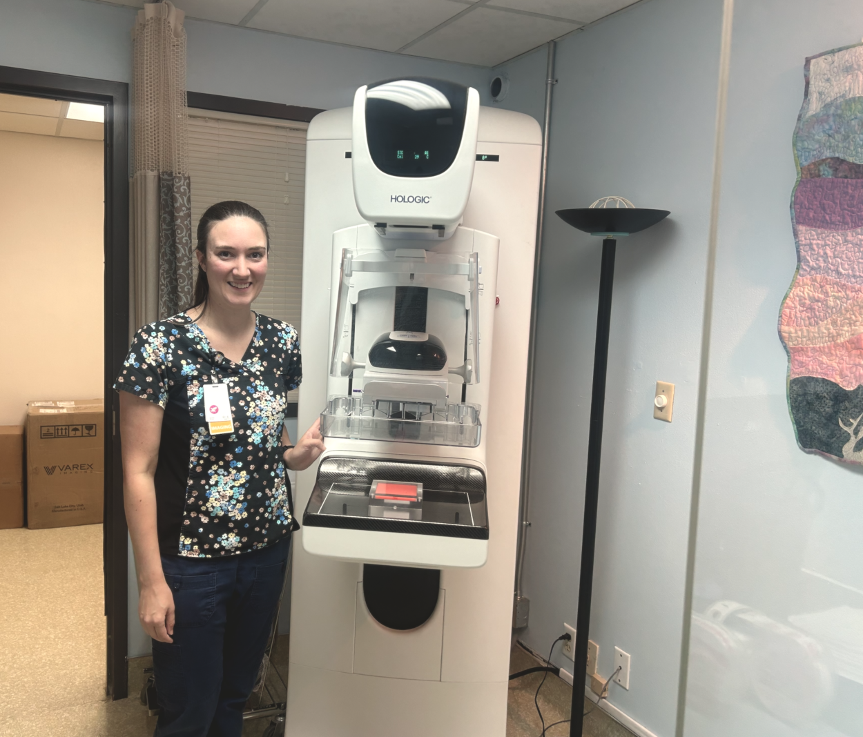
Petersburg Medical Center has a new mammography machine that could catch breast cancer and other abnormalities earlier, and with more accuracy. Hospital staff say it produces better pictures, but also happier patients: the new tech might lessen the discomfort of breast exams.
Sonja Paul is Petersburg Medical Center’s radiology manager. The hospital’s new mammography machine, a Hologic 3D Unit, arrived in early March, and part of her job is to calibrate it every week. She gave me a little demonstration.
“A patient’s breast goes on this part of the machine — which is basically what my image receptor is,” said Paul, as she gestured towards a horizontal panel on the machine, positioned about waist-high. “The compression pedal comes down against the breast tissue. What I am looking for is not over-compressed, but enough to make the breast tissue taut and tight. So you’re not going to move – it’s going to spread that tissue out.”
She placed what’s called a “phantom slide” over the compression pedal, where a patient’s breast would ordinarily go. It’s a block of pink-tinted acrylic that has little bits and pieces hidden inside that aren’t visible to the naked eye. But if the machine is working correctly, those flecks should appear in a radiographic image. She takes pictures of phantom slides every week to make sure everything is in working order.
“Once you’re in your position, I come back to my machine,” said Paul. “It’s got all of the parameters set, and [while] it takes an exposure, I ask you to hold your breath and then the machine starts. Once it’s done, you automatically get released!”
Then she takes me behind a giant computer screen in the back of the room to show me what her giant camera got.
“So, my control panel back here shows me the image that was just created,” said Paul. “I can look through those 3D projections, just to make sure that I’m giving the radiologist what they need.”
If Paul were taking pictures of a real patient, she’d send the images off to an out-of-state radiologist through a secure electronic program.
The whole exam takes just a few minutes. And Paul said the process isn’t as scary as a lot of Paul’s new patients expect.
“It is intimidating,” said Paul. “My advice is just… I mean, I hate to say it, but just come in and have it done! It’s a lot better than what you mentally picture. I’ve also heard it from my first-time exam ladies: ‘That wasn’t as bad as I anticipated!'”
She said the new machine might even make the whole experience a bit more comfortable.
“Many women that are here year after year say it’s not the same mammogram that women have always [spoken] of,” said Paul. “There are little things that have been changed with it that are supposed to help with comfort — it’s got flexibility to the compression paddle, which is supposed to help. It’s got some more rounded corners, things like that. Breast exams and mammograms, they’re uncomfortable, but they shouldn’t hurt! I don’t want that to be a barrier between women receiving their exams.”
But most importantly, she said the new machine gives a much more thorough exam. And that could save lives.
The radiology department’s waiting list for mammograms is short, at the moment. Paul said they can get a new patient in for an exam within a week. Paul reminds women in vulnerable categories to get their breasts checked on schedule. That means something different to different healthcare groups — from the Center for Disease Control, to the American Cancer Society, to the American College of Radiology. But they generally agree that women with an average risk of breast cancer should get their first mammogram around the age of 40, and then about every year after that.
Paul said exam frequency is important for catching early signs of breast cancer. But getting screened with the right equipment is another important factor in positive outcomes: “The better screening, the better the prognosis,” she said.


